Common Types of Echocardiograms

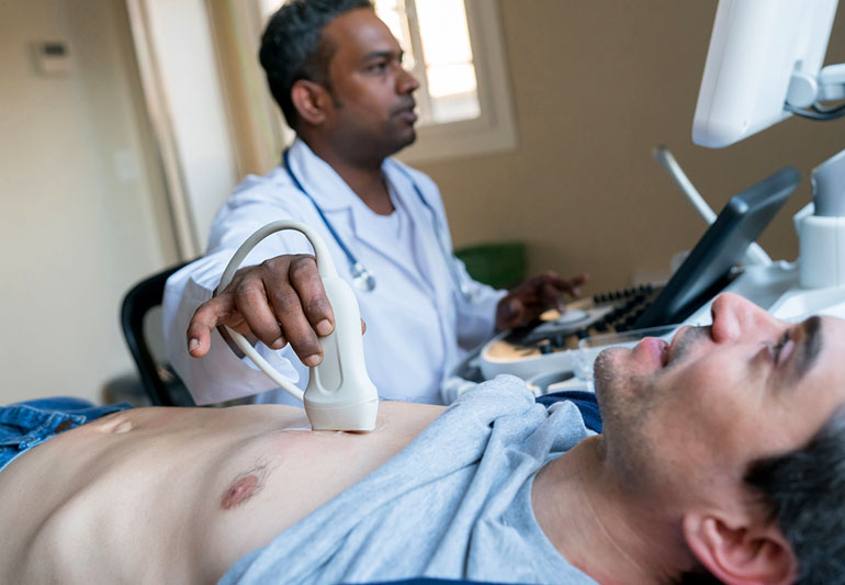
The echocardiogram is a test used to examine the heart and determine if it is normal and healthy. The echocardiogram is a non-invasive diagnostic procedure that uses sound waves to create pictures of the heart and its surrounding structures. A cardiologist can evaluate patients with chest pain, chronic heart failure or other heart abnormalities using an echocardiogram Philadelphia.
There are several different types of echocardiograms used in clinical practice. Each type may be appropriate for a specific patient or situation. Researchers also use echocardiograms to study health problems that involve the heart, such as congenital heart disease and valvular disease.
The most common types of echocardiograms are:
Holter monitor
A Holter monitor is a test that records your heart rate, rhythm and activity for 24 hours. It is often done for people with chest pain or shortness of breath that may be due to an irregular heartbeat. Also, people at risk for heart failure or those who want to monitor changes over time in their heart rhythm or function should undergo this test.
Cardiac resynchronization therapy (CRT)
This procedure involves implanting a pacemaker and, or defibrillator into your heart. It is an alternative treatment option for people with life-threatening arrhythmias caused by abnormal electrical activity in the heart muscle.
Color-coded Doppler echocardiogram
A color-coded Doppler echocardiogram requires two machines – one for imaging and one for real-time measurement of blood flow velocity. It gives doctors a complete picture of how well blood flows through various parts of the heart muscle, helping them determine if there are any problems with blood supply or blockages in these areas.
Left ventricular (LV) echocardiogram
Left ventricular (LV) is a type of echocardiogram doctors use to look at the left side of the heart and can be used to diagnose cardiac problems or assess LV function. It may also be used with other tests to provide insight into certain diseases or conditions, such as aortic stenosis, valvular disease, hypertrophic cardiomyopathy and mitral valve prolapse.
Maternal echocardiogram
A maternal echocardiogram, also called a maternal echo, is done on pregnant women to check for structural problems in their hearts or valves and determine if there are any problems with the fetus’s heart function. An ultrasound probe is placed over the mother’s abdomen during this test, and sound waves are sent into her uterus with an ultrasound machine. The echoes are then received by another machine and translated into images that show how well the heart is functioning and how well blood flows through it during pregnancy.
An echocardiogram is a valuable tool for evaluating cardiac function, such as how well the heart pumps blood or whether there are any problems with valves or other structures inside your heart. You might have a family history of heart disease or an account of heart disease in your immediate family. In that case, it is essential to consult a cardiologist to rule out a heart attack or other severe cardiac problems. Contact Corrielus Cardiology today to schedule an appointment with a cardiologist so you can learn more about echocardiograms.


